Researchers develop tiny nanoSABERs to aid battle against cancer
New technology allows clinicians to visualize tumors in their entirety, which could improve cancer imaging and outcomes
When Jedi Knights need to vanquish an enemy, they whip out their trusty lightsabers. In the future, thanks to Johns Hopkins researchers, doctors seeking to crush cancer may wield minuscule molecular nanoSABERs that allow them to look at tumors in ways never before possible.
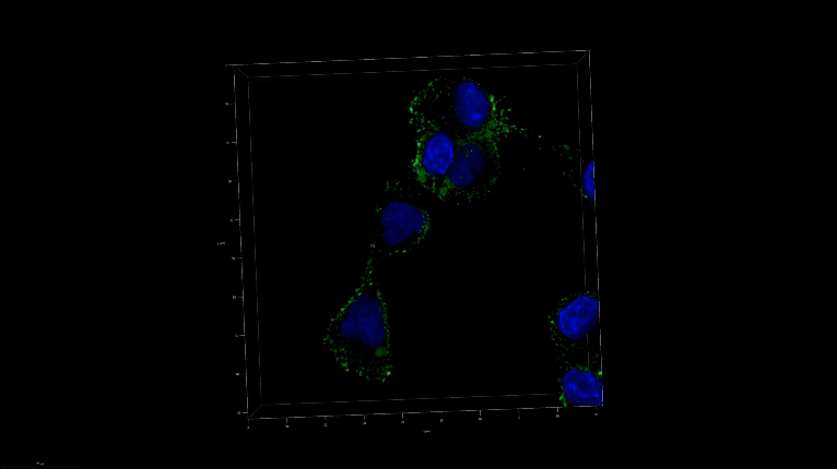
Inspired by the process cells use to assemble proteins, a team led by two researchers—Ishan Barman at the university’s Whiting School of Engineering and Jeff W. Bulte, a professor of radiology and radiological science at the School of Medicine who is also affiliated with JHU’s Institute for NanoBioTechnology—has created infinitesimal probes that light up when they encounter certain enzymes found in cancer cells. The ability to visualize tumors in their entirety—and early—could significantly enhance cancer imaging, inform treatment options, and improve patient outcomes.
“This could be a game changer for cancer treatment,” said Barman, an associate professor of mechanical engineering at the Whiting School, of the self-assembling biorthogonal enzyme recognition (nanoSABER) probes. The team’s results appear in Advanced Science.
Currently, tissue biopsies are the gold standard for detecting most cancers, though they can be inexact and even miss parts of tumors lurking in the margins. The Johns Hopkins team’s approach could solve that problem, allowing clinicians to visualize cancerous activity across entire tumors, providing insights into their possible aggressiveness.
Enzymes, especially legumain, play a leading role in the development and progression of cancer.
The team’s new tool assembles itself in the presence of these cancer-related enzymes and emits a signal that can then be picked up by Raman spectroscopy, a visualization technique that analyzes molecular vibrations to identify and characterize substances. This allows the probes to pinpoint cancer cells accurately.
The Johns Hopkins team says its method also could allow clinicians to more accurately monitor the accumulation of cancer drugs in tumors during treatment, providing an indication of how well those treatments are working.
“The probes’ ability to offer a clear look at the molecular, cellular, and tissue levels provides a comprehensive perspective,” said lead author Swati Tanwar, a post-doctoral fellow in mechanical engineering. “It is imperative to understand what is really happening at the tumor margins to ensure complete cancer removal and minimize the chances of recurrence.”
Study co-authors at Johns Hopkins include Behnaz Ghaemi, Piyush Raj, Aruna Singh, Lintong Wu, Dian R. Arifin, and Michael T. McMahon. The team also included Yue Yuan of the University of Science and Technology of China.
Story by Lisa Ercolano