New Magnetic Particle Imager Offers New Opportunities in Molecular and Cellular Imaging Research
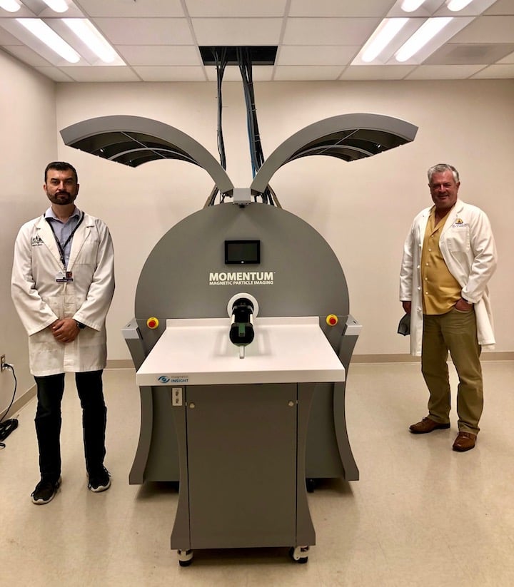
Above image: Adnan Bibic (left) and Jeff Bulte (right) with the magnetic particle imaging scanner.
A magnetic particle imaging (MPI) scanner, a new imaging modality, was installed at the Hugo W. Moser Research Institute at Kennedy Krieger Institute with the help of Jeff W.M. Bulte, PhD, a specialist in molecular and cellular imaging. Bulte, an affiliate faculty member at the INBT, a professor of radiology and radiological science, and director of cellular imaging in the Institute for Cell Engineering at the Johns Hopkins University School of Medicine, obtained the scanner through an NIH S10 shared instrumentation grant. The facility is supported through a collaborative effort between Kennedy Krieger and the Johns Hopkins Department of Radiology. The new instrument is the first of its kind on the East Coast and allows for cutting-edge molecular and cellular imaging research.
MPI is ultrasensitive and can detect just a few nanograms of magnetic material. Nanoparticles made of magnetic materials, such as cobalt, nickel or iron, are widely used in many applications ranging from aerospace to biomedicine. Many magnetic nanoparticles are unsuitable for biomedical applications because of toxicity or poor biocompatibility. However, magnetic iron oxide nanoparticles have become important in biomedical applications with several iron oxide nanoparticle compounds approved for magnetic resonance imaging (MRI) contrast and therapy. MPI is the only technology that enables direct visualization and quantitation of magnetic nanoparticles because the technique directly measures the magnetic signature of the nanoparticles. Thus, with MPI, a magnetic iron oxide nanoparticle is a tracer, not a contrast agent as it is for an MRI. This allows for dose-response studies, such as quantifying how many magnetically labeled stem cells or CAR T cells are at a target site, and how these cell numbers correlate to therapeutic outcomes. The device also has theranostic applications in gene and drug delivery as it can tell researchers how many magnetic nanoparticles need to accumulate at the target site to induce the desired gene expression and anti-tumor response. Furthermore, the technology can leverage nanoparticles to image functional events.
As part of the shared instrumentation grant, Bulte is partnering with 11 investigators from several departments across Johns Hopkins and the Kennedy Krieger Institute to develop applications using the MPI scanner. The proposed projects will be interdisciplinary and range from materials engineering, biomedical engineering, immunology, diabetes, gene therapy, and drug delivery to molecular imaging. Most of these projects are highly relevant to the precision medicine initiative. Bulte is also creating an MPI user group to discuss potential applications, tracer development, and advanced training. The group plans to hold regular seminars to present the progress of research projects.
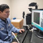
The MPI scanner was installed at the end of May and is managed by Adnan Bibic, PhD, as part of the pre-clinical imaging facility of the F.M. Kirby Center for functional brain imaging, at the Kennedy Krieger Institute, led by Peter van Zijl, PhD.
If you are interested in joining the new MPI user group initiative, email Jeff Bulte at jwmbulte@mri.jhu.edu.