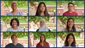
Every summer for 12 years, the INBT has welcomed undergraduate students to the Nanotechnology for Biology and Bioengineering Research Experience for Undergraduates (REU) program. Students spend 10-weeks with INBT faculty and mentors heavily engaged in research projects ranging from developing cancer therapies and diagnostic tools to using regenerative engineering to heal the body. They also participate in professional development training, networking activities, and explore Baltimore and other surrounding cities. We welcome you to join us to see presentations by our 2022 summer students as they showcase their research projects.
This event is hybrid. Space is limited in Malone Hall G33/G35 to 35 people. If space is unavailable we ask you to join by Zoom.
Zoom information
https://wse.zoom.us/j/94977263610?pwd=WFRNRU1TbEFhclBOdkxvdkxwNGI0Zz09
Meeting ID: 949 7726 3610
Passcode: 146035

FastForward U’s accelerator programming is an opportunity for student teams from across the University to work collaboratively to make progress on their ventures. These engaging, cross-disciplinary initiatives build skills, grow networks, and connect founders with other entrepreneurial students.
Teams are grouped by stage to allow students to learn together at a pace that makes sense for where they are on their entrepreneurial journey. Spark and Fuel tracks include a stipend and the chance to win additional funds at Demo Days.
Learn more about each track.
“The Discovery of Viscosity Sensor that Facilitates the Counterintuitive Acceleration of Migrating Cells in Highly Viscous Fluids”
Yun Chen is an assistant professor of mechanical engineering at Johns Hopkins University. Her research is focused on developing tools to measure key parameters in mechanobiology, understanding the fundamental biophysical mechanisms that contribute to diseases, and applying knowledge gained from basic mechanobiology research to clinical applications. While a vast amount of effort has been invested in characterizing the biophysical properties in diseased cells and tissues, most of these efforts are limited to measuring the stiffness, diffusion, and viscosity of samples. Those properties are regarded as consequences of the diseases, rather than the causes. The abnormal biophysical traits can be the causes of the diseases, and her research has been dedicated to uncovering this commonly overlooked causality. Similarly, the unusual biophysical properties associated with diseases have been exploited as diagnosis tools, but few treatments, if any, employ biophysical principles to correct the errant biological processes known as pathology. Chen’s research group has been making significant progress on these uncharted territories. Their goal is to understand how altered biophysics in biological systems contribute to pathological processes in order to develop treatments for diseases. Their efforts include developing measurement tools to quantitatively characterize biophysical phenomena, such as axial stiffness of twisted DNA strands, differential force generation profiles and viscoelasticity of cancer cells compared to their normal counterparts, and identifying the underlying mechanisms for such differences, which can be exploited for disease diagnosis and treatment.
Extracellular fluid (ECF) is a critical component of the body. Cells are surrounded by and move through biological fluids that span orders of magnitudes of viscosity in vivo, including mucus, saliva, blood, and synovial fluid, among others. Interstitial fluid in the tumor microenvironment is viscous, ascites in cancer patients is highly viscous, and the mucus of patients with cystic fibrosis is highly viscous. Elevated viscosity in the tumor microenvironment and in ascites can increase the rate of cancer cell motility and promote metastasis. Elevated viscosity in mucus can inappropriately increase the migration of fibroblasts to airway wounds incurred in patients with cystic fibrosis, resulting in the worsening of fibrosis. Increases in ECF viscosity are also associated with aging and many other diseases. Despite the profound implications of ECF viscosity, our understanding of the mechanosignaling pathways that allow cells to respond to viscosity changes and the underlying mechanism leading to increased cell speeds is very limited. To gain more insights, we used bio-compatible polymers to mimic viscous ECF, aims to fill this knowledge void. We conducted detailed characterization of the cellular responses to viscosity – from the time point immediately after viscosity is increased to hours afterwards, and from single molecule force measurement to dynamic 3D cellular morphology profiling. We observed that cells immersed in highly viscous medium, which had a consistency similar to that of honey, drastically changed morphology and began moving nearly twice as fast. Step by step, we dissected the molecular cascade leading to the cell speed increase in response to elevated viscosity. Combining numerical simulation and experimental data, we showed that membrane ruffling, a common feature of adherent cells, acts in effect as a sensor of ECF viscosity, probing the hydraulic resistance of the surrounding fluid and triggering adaptive responses. In summary, we demonstrate for the first time that a universal viscosity sensing mechanism exists in adherent cells to actively probe and adapt to changes in the viscosity of the microenvironment. The physical interplay between mechanical forces that power membrane ruffling and the counteracting hydraulic resistance is at the heart of this sensing mechanism.

Uniquely among mammalian organs, skin is capable of dramatic size changes in adulthood, yet the mechanisms underlying this striking capacity are unclear. The Reddy lab recently developed a method to induce controlled skin growth in genetically tractable mice. Using machine learning-guided three dimensional tissue reconstruction, they discovered that skin growth is driven by proliferation of the epidermis in response to mechanical tension, with more limited changes in dermal and subdermal compartments. Epidermal growth is in turn achieved through preferential activation and differentiation of Lgr6+ stem cells of the epidermis, controlled in part by the Hippo pathway. Single-cell RNA sequencing uncovered further changes in mechanosensitive and metabolic pathways underlying growth control in skin. These studies point to therapeutic strategies to enhance skin growth and establish a platform for understanding organ size dynamics in adult mammals.
Those who cannot attend in person can watch on Zoom.
About the speaker
Dr. Sashank Reddy completed his undergraduate studies at Johns Hopkins as a Beneficial Hodson Scholar, followed by MDPhD studies at Harvard Medical School and MIT under the auspices of the NIH Medical Scientist Training Program. Following his clinical training at the Johns Hopkins University School of Medicine, Dr. Reddy joined the faculty in 2019. His NIH-funded laboratory studies developmental biology and regenerative medicine with a particular focus on soft tissues. Dr. Reddy is also an accomplished biomedical innovator and a founder of venture-backed companies. In his role at INBT, Dr. Reddy works to grow the scientific and translational excellence of the Institute.
From Bench to Bedside: Translation of a Novel PLGA Nanoparticle Delivery System for Tolerogenic Therapy of Immune-Medicated Diseases
Seminar Take-Home Points
- Tolerance induction using antigenencapsulating PLG nanoparticles (Ag-PLG) recapitulates how self-tolerance is induced and maintained in the hematopoietic system.
- Ag-PLG uptake by splenic marginal zone and liver APCs via the MARCO scavenger receptor confers a tolerogenic phenotype and induces activation of CD4+Foxp3+, CD4+ Tr1, andCD8+CD122+ regulatory T cells which regulate effector T cell responses in a IL-10-dependentmanner.
- Gliadin-encapsulating PLG nanoparticles have proven efficacious in a phase 2 double-blind, placebo-controlled trial in celiac disease patients significantly reducing the gliadin-specific T cell response and preventing intestinal damage upon gluten challenge.
- Future disease indications under clinical development include multiple sclerosis (MS),neuromyelitis optica (NMO), and peanut allergy.
About the speaker
Dr. Stephen Miller is the Judy E. Gugenheim Research Professor Emeritus of MicrobiologyImmunology at Northwestern University Feinberg School of Medicine in Chicago. He received his Ph.D. in 1975 from the Pennsylvania State University and did postdoctoral training at the University of Colorado Health Sciences Center before joining the faculty at Northwestern in 1981 where he founded and served as Director of the Northwestern University Interdepartmental Immunobiology Center from 1992-2021. Dr. Miller is internationally recognized for his research on pathogenesis and regulation of autoimmune diseases. His current work is geared towards understanding the cellular and molecular mechanisms of T cell tolerance and translating the use of antigen-encapsulating biodegradable PLG nanoparticles for the treatment of other human immunemediated diseases including autoimmunity, allergy, protein and gene replacement therapy, and tissue/organ transplantation.
All are welcome to a seminar with guest speaker Gregg Duncan and his presentation on, “Mucus Gels and Innate Lung Defense.” This is a hybrid event. Guests are welcome to come in-person in Hodson Hall 210 on the Johns Hopkins Homewood campus or by Zoom.
Zoom link and passcode: 530803
Mucus is a biological gel within the lung designed to behave like an “escalator” with the ability to capture potentially harmful inhaled materials (e.g. pathogens, particulates) and carry these materials via mucociliary clearance up to the throat to be swallowed and sterilized. A breakdown in lung mucus barrier function can lead to increased infections by respiratory viruses, such as influenza, rhinovirus, and coronaviruses, as they are not effectively removed from the airway. For these seasonal and emerging human viral pathogens, it is important to understand the mechanisms through which viral particles avoid adhesion to the mucus barrier and transport to the underlying epithelium to cause infection. To examine this, we measured influenza A virus and nanoparticle diffusion in mucus from human donors using high-speed fluorescent video microscopy and multiple particle tracking. Through these measurements, we can directly determine binding affinity and mode of adhesion for influenza A and other respiratory viruses in 3D human mucus matrices. MUC5B and MUC5AC are large, gel-forming mucins that assemble to form airway mucus gels. However due to the lack of appropriate models, it is not yet fully understood how MUC5B and MUC5AC individually or synergistically contribute to the biological function of mucus. To understand their unique roles in respiratory health, I will also discuss our studies on the rheological properties and transport function of mucus in human airway tissue cultures genetically engineered to secrete either MUC5B or MUC5AC. These bioengineered models provide key insights on how MUC5B and MUC5AC work in concert to enable host mucosal barrier function providing a highly valuable means to understand their roles in health and disease.
Speaker Bio: Gregg Duncan earned his Ph.D. in chemical engineering under the guidance of Michael Bevan at Johns Hopkins University. He then completed his postdoctoral training at Johns Hopkins School of Medicine in the Center for Nanomedicine directed by Justin Hanes. Dr. Duncan is currently an Assistant Professor in the Fischell Department of Bioengineering at the University of Maryland. Dr. Duncan leads the Respiratory Nano Bioengineering (RnB) lab, which aims to understand airway micro-physiology in health and disease to engineer new therapeutic strategies for obstructive lung diseases such as asthma, chronic obstructive pulmonary disease, and cystic fibrosis. Dr. Duncan is the recipient of several honors and awards including the Burroughs Wellcome Fund Career Award at the Scientific Interface, BMES Rita Schaffer Young Investigator Award, the CMBE Young Innovator Award, and the NSF CAREER Award
Advancing Biological Imaging and Sensing Using Quantum Technologies with Warwick Bowen.
Quantum technologies can exponentially accelerate computer simulations and detect signals that would be invisible to other technologies. This provides the potential for wide impact across the biosciences: better modelling of biochemical processes, and better imaging of biological systems. In this talk I will provide an overview of this potential, and how it could create a new field of quantum biotechnology. As an illustrative example, I will then introduce work in my laboratory that uses quantum correlated light to enhance bioimaging. In that work, we demonstrate for the first time that quantum correlations can be used to evade photochemical intrusion on the biological specimen, and therefore to observe biological structures that would be otherwise inaccessible. We achieve this in a coherent Raman microscope, providing chemically-specific information about the cell. Our results represents the first demonstration of absolute quantum advantage in biological microscopy, and we hope will open the door to a bright future for quantum bioimaging.
Probing and Attacking the Cancer Surfacome with Jim Wells, PhD
The cell surface proteome(surfaceome)is a major hub for cellular communication and a primary source of drug targets, especially for biologics. My lab is interested in developing proteomic means to probe how the surfaceome changes in health and disease, especially cancer. Such changes involve alteration in protein expression and post-translational modifications such as proteolysis. I’ll describe new engineered tools we have built to probe the surfaceome changes that occur when oncogenes are expressed in isogenic cell lines to identify targets of interest. We then target proteins either upregulated, proteolyzed or both with recombinant antibodies derived by phage display to be used as validation tools and potential therapeutic leads
Jim received his BA from University of California at Berkeley, PhD from Washington State University (with Ralph Yount), and post-doc at Stanford (with George Stark), prior to joining Genentech, then Sunesis Pharmaceuticals, and finally UCSF. Wells’ group pioneered the engineering of proteins, antibodies, and small molecules that target catalytic, allosteric, and protein-protein interaction sites; and technologies including protein phage display, alanine-scanning, engineered proteases, bioconjugations, N-terminomics, disulfide “tethering”, and more recently an industrialized recombinant antibody production pipeline for the proteome. His team was integral to several protein products including Somavert for acromegaly, Avastin for cancer, Lifitegrast for dry eye disease, and engineered proteases sold by Pfizer, Genentech, Shire and Genencor, respectively. He is an elected member of the US National Academy of Science, American Association of Arts and Science, and the National Academy of Inventors.
This is a virtual event on Zoom. Click here to get the link.

The Advances in Immunoengineering: Fundamentals and Cutting Edge Advances workshop is hosted by Johns Hopkins Translational Immunoengineering. The workshop meets twice a week for three weeks and participants are eligible for CME credit. The workshop is also offered as a two-credit course to Johns Hopkins students
The immunoengineering field is transforming cancer, autoimmunity, regeneration, and transplantation treatments by combining the diverse and complex fields of engineering and immunology. There is a significant need to train engineers in immunology and immunologists in quantitative engineering techniques. Moreover, there is a need to bridge basic immunological discoveries with advances in clinical application. This workshop will review immune system fundamentals and components, engineering strategies to modulate the immune system, and clinical applications.
After attending this workshop, the learner will demonstrate the ability to:
– Review the fundamentals and recent discoveries in the function of the immune system.
– Identify engineering strategies to manipulate the immune system.
– Describe the clinical applications of immunoengineering.
The full schedule, speakers, topics, and registration information are available on JH-TIE’s website.


