Landmark physical characterization of cancer cells completed

An enormous collaborative effort between a multitude of academic and research centers has characterized numerous physical and mechanical properties on one identical human cancer cell line. Their two-year cooperative study, published online in the April 26, 2013 journal Science Reports, reveals the persistent and agile nature of human cancer cells as compared to noncancerous cells. It also represents a major shift in the way scientific research can be accomplished.
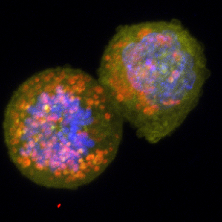
In the image above:
Human breast cancer cells like these were used in the study. (Image created by Shyam Khatau/ Wirtz Lab)
The research, which was conducted by 12 federally funded Physical Sciences-Oncology Centers (PS-OC) sponsored by the National Cancer Institute, is a systematic comparison of metastatic human breast-cancer cells to nonmetastatic breast cells that reveals dramatic differences between the two cell lines in their mechanics, migration, oxygen response, protein production and ability to stick to surfaces. They have also discovered new insights into how human cells make the transition from nonmalignant to metastatic, a process that is not well understood.
Denis Wirtz, a Johns Hopkins professor of chemical and biomolecular engineering with joint appointments in pathology and oncology who is the corresponding author on the study, remarked that the work adds a tremendous amount of information about the physical nature of cancer cells. “For the first time ever, scientists got together and have created THE phenotypic signature of cancer” Wirtz said. “Yes, it was just one metastatic cell line, and it will require validation with many other cell lines. But we now have an extremely rich signature containing many parameters that are distinct when looking at metastatic and nonmetastatic cells.”
Wirtz, who directs the Johns Hopkins Physical Sciences-Oncology Center, also noted the unique way in which this work was conducted: all centers used the same human cell line for their studies, which makes the quality of the results unparalleled. And, since human and not animal cells were used, the findings are immediately relevant to the development of drugs for the treatment of human disease.
“Cancer cells may nominally be derived from the same patient, but in actuality they will be quite different because cells drift genetically over just a few passages,” Wirtz said. “This makes any measurement on them from different labs like comparing apples and oranges.” In this study, however, the genetic integrity of the cell lines were safeguarded by limiting the number times the original cell cultures could be regrown before they were discarded.
The nationwide PS-OC brings together researchers from physics, engineering, computer science, cancer biology and chemistry to solve problems in cancer, said Nastaran Zahir Kuhn, PS-OC program manager at the National Cancer Institute.
“The PS-OC program aims to bring physical sciences tools and perspectives into cancer research,” Kuhn said. “The results of this study demonstrate the utility of such an approach, particularly when studies are conducted in a standardized manner from the beginning.”
For the nationwide project, nearly 100 investigators from 20 institutions and laboratories conducted their experiments using the same two cell lines, reagents and protocols to assure that results could be compared. The experimental methods ranged from physical measurements of how the cells push on surrounding cells to measurements of gene and protein expression.
“Roughly 20 techniques were used to study the cell lines, enabling identification of a number of unique relationships between observations,” Kuhn said.
Wirtz added that it would have been logistically impossible for a single institution to employ all of these different techniques and to measure all of these different parameters on just one identical cell line. That means that this work accomplished in just two years what might have otherwise taken ten, he said.
The Johns Hopkins PS-OC made specific contributions to this work. Using particle-tracking microrheology, in which nanospheres are embedded in the cell’s cytoplasm and random cell movement is visually monitored, they measured the mechanical properties of cancerous versus noncancerous cells. They found that highly metastatic breast cancer cells were mechanically softer and more compliant than cells of less metastatic potential.
Using 3D cell culturing techniques, they analyzed the spontaneous migratory potential (that is, migration without the stimulus of any chemical signal) of cancerous versus noncancerous cells. They also analyzed the extracellular matrix molecules that were deposited by the two cell lines and found that cancerous cells deposited more hyaluronic acid (HA). The HA, in turn, affects motility, polarization and differentiation of cells. Finally, the Hopkins team measured the level of expression of CD44, a cell surface receptor that recognizes HA, and found that metastatic cells express more CD44.
The next steps, Wirtz said, would be to validate these results using other metastatic cell lines. To read the paper, which is published in an open access journal, follow this link: http://www.nature.com/srep/2013/130422/srep01449/full/srep01449.html
Excerpts from original press release by Princeton science writer Morgan Kelly were used.
Latest Posts
-
 Q&A with PSON Intern Jocelyn Hsu
August 19, 2021
-
 Start Up Founders from Johns Hopkins Aim to Stop Spread of Cancer
August 3, 2021
-
 Protein Appears to Prevent Tumor Cells from Spreading Via Blood Vessels
July 15, 2021


